 VEYLO
VEYLOHow Veylo compares
Veylo, clinical 3D systems and AI apps: how each one works.
Clinics use VECTRA, Crisalix, LifeViz and 3dMD; AI apps turn a selfie into an edited photo. Veylo is an iPhone app that visualizes treatments in real units. Here is how each works, what studies say, and where Veylo still has to prove itself.
01Overview
Four ways to show a facial change, each built for a different job.
- Clinic 3D cameras
VECTRA, LifeViz, 3dMD
Several calibrated cameras photograph the face at the same moment (stereophotogrammetry) and compute a 3D surface. VECTRA XT captures in 3.5 ms, the handheld H2 in 2.0 ms[7, 8]. For a simulation, a clinician shapes the 3D face with sculpting tools[7].
- Photos to 3D, online
Crisalix
Three ordinary photos are uploaded and turned into a 3D model online[9]. Clinics subscribe; patients can buy a one-off simulation from €24[9, 10]. Crisalix also co-developed FACE by Galderma, an augmented-reality tool for injectables[11].
- Consumer AI apps
Generative photo editors
A selfie plus a prompt or preset goes to an image model, which paints a new photo. The result looks photographic, but the model decides how much changes and where; a 2026 study of commercial editors found nose and jaw edits that also changed other parts of the face[12].
- Veylo
Phone depth and real units
The iPhone TrueDepth camera captures depth from several angles at home. You set a treatment in ml, units, mm or an implant model and size, and the app deforms your own scan and photo by an amount derived from published studies. An optional realistic view renders it as a photo.
02How Veylo works
How Veylo works, step by step.
 1
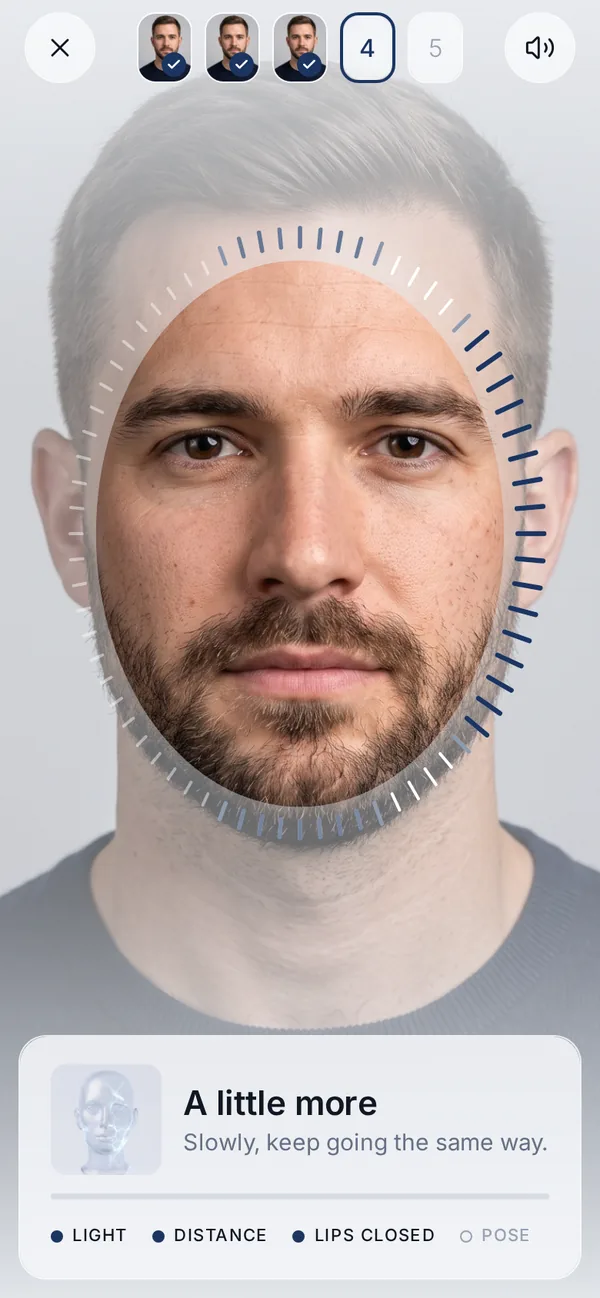
1Capture with TrueDepth
Five guided angles with the depth camera behind Face ID, in about 40 seconds.
 2
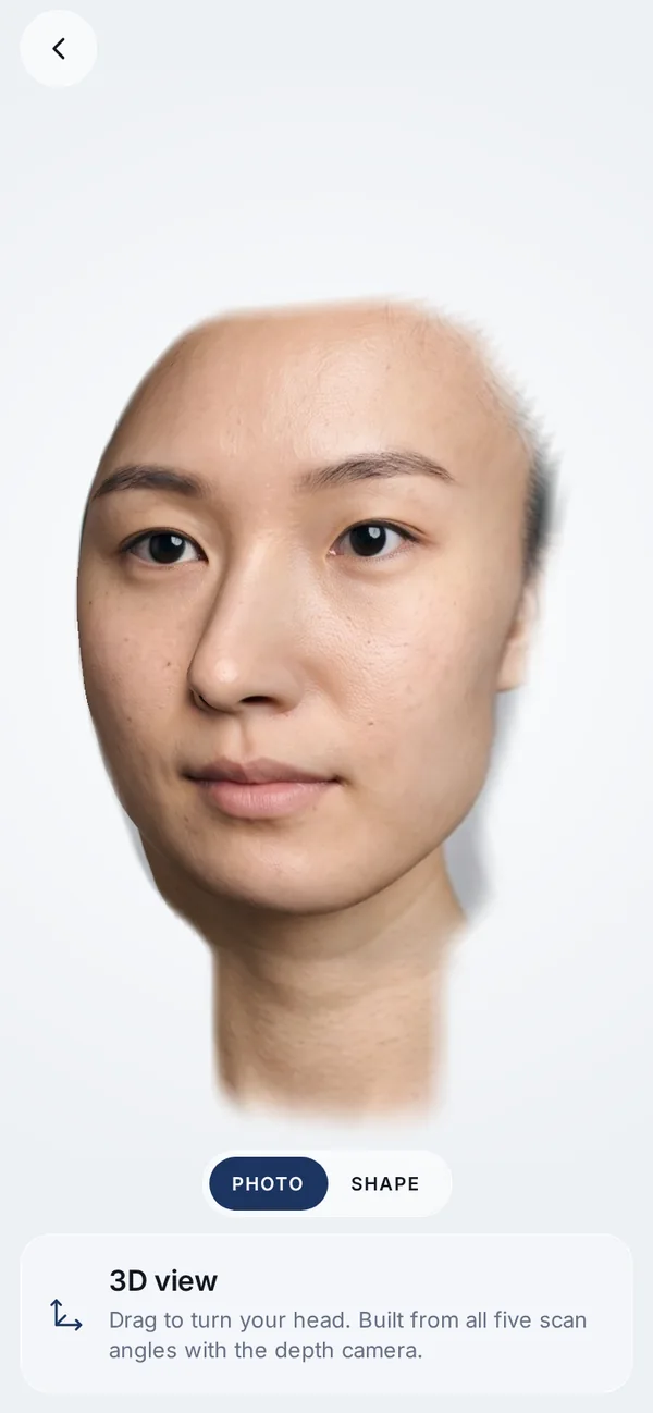
2Fuse the depth
The depth from each angle is aligned and merged into one 3D surface.
How, with sources
 3
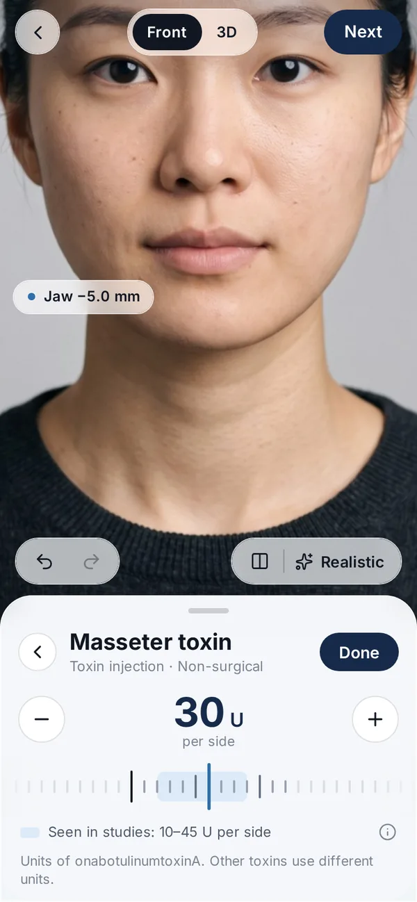
3Deform in real units
You set ml, units, mm or an implant size; the engine turns it into soft-tissue movement.
How, with sources
You set a treatment in the unit a clinic uses. The engine turns it into soft-tissue movement with ratios from studies: for example, the chin’s soft tissue moved 83–87% of an implant’s size[15] and about 0.9 mm per mm of genioplasty advancement[16]. Automated tests check that other face areas move by exactly zero and that the same settings always give the same image.
 4
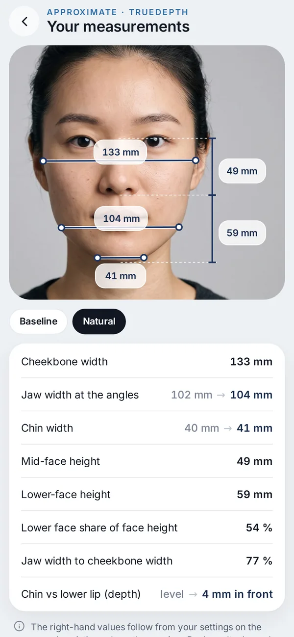
4Read approximate mm
Before → after readouts come from the same numbers as the picture.
How, with sources
Before → after readouts such as jaw width or chin projection come from the same numbers. They describe what the visualization changed, not what a treatment will measure.
 5
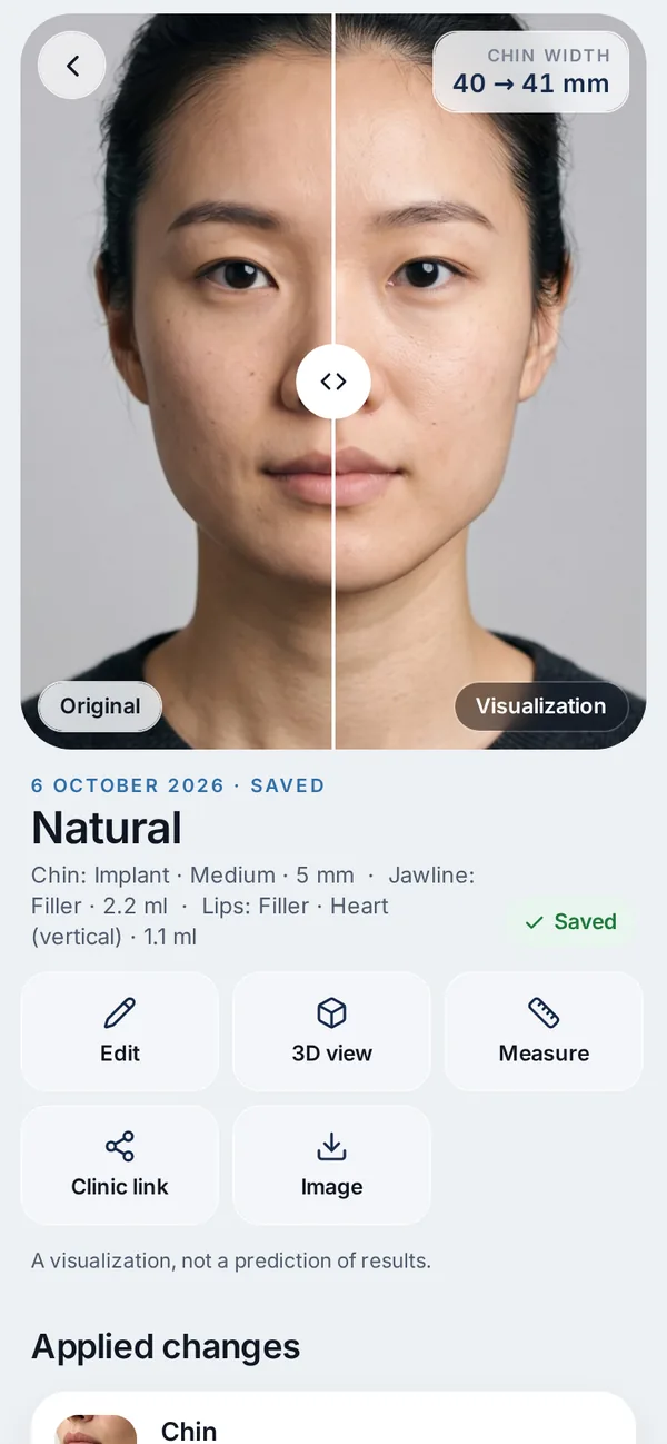
5Optional realistic view
On request and after consent, a photographic render of the edit.
How, with sources
Faces in app screens are synthetic.
03Side by side
Side by side, from public pages and published studies.
| Veylo | VECTRA (Canfield) | Crisalix | LifeViz (QuantifiCare) | 3dMD | Consumer AI apps | |
|---|---|---|---|---|---|---|
| Capture | iPhone TrueDepth depth and photos; 5 guided angles at home, about 40 s | Stereophotogrammetry; XT: 1.2 mm mesh, 3.5 ms capture; H2 handheld: 2.0 ms[7, 8] | 3 photos from a phone or camera, 3D model built online[9] | Portable stereo camera: 3 photos stitched, light pointers for repeatable positioning[19]; Infinity on a motorised platform[20] | Multi-camera 3D and 4D capture for research and healthcare[21] | One 2D selfie |
| Published accuracy | Not validated yet (plan below). Other TrueDepth apps vs VECTRA: 0.44 mm and 1.1 mm[1, 2] | Review of 10 validation studies: accurate and reproducible, minor errors around the mouth[22]; H1 error bound 0.40 mm[23] | Detected 0.5–4 cc volume changes in a five-device comparison[24]; we found no face-surface distance study | Same five-device comparison: good accuracy for 0.5–4 cc changes[24] | 0.36 mm vs calipers on a mannequin[25]; error bound 0.44 mm[23] | No geometric accuracy data; one GAN study measured realism only[26] |
| How the simulation is made | Treatment + amount → soft-tissue change from published ratios; other areas fenced off | Clinician shapes the face with constrained and unconstrained tools; breast simulation uses implant catalogues and a gravity model[7] | 3D simulation by the clinic, or by the patient from home[9] | Real-time simulation tools; automatic breast implant recommendations[20] | Capture and measurement; no aesthetic simulator listed on its site[21] | Image model edits the photo from a prompt or preset |
| Units in → out | In: ml, units, mm, implant model and size, nose or lip style. Out: approximate mm | In: a sculpted shape. Out: distances, colour distance map, volume difference in one click[7] | Not stated per ml on the public pages we reviewed | Measurements on the 3D image[20] | Measurements on the 3D image | No units |
| Filler change over time | Shows a settled result; a timeline view based on published curves is in testing | Measures change between visits (Markerless Tracking)[8] | Not described on the public pages we reviewed | Follow-up comparisons between visits[20] | Repeat captures possible; no aesthetic timeline listed | No |
| Price and access | Free: scan, editor, 3D, measurements, 1 realistic view. Premium $39.99 a year or $9.99 a month. iPhone with Face ID | Clinic hardware, price on request; patients see results in the clinic or in the ViewMyConsult portal[27] | Clinic plans on request (Gold: 60 patients a year; Platinum: unlimited, fair use); patients from €24 one-off[10, 9] | Clinic hardware[19] | Research and clinical hardware[21] | Usually a free download with in-app purchases[28] |
| Where face data is kept | Scans, depth and measurements stay on the iPhone; the realistic view sends the photo only after consent; nothing goes to analytics[17] | Clinic system; password-protected web portal for patients[27] | Photos uploaded to a Crisalix account[9] | Clinic system | Clinic or research system | Photo processed by the app’s cloud model; varies by app |
| Who runs it | The person themselves; can share an encrypted link with a clinic | A clinician, in the clinic | The clinic, or the patient from home | A clinician | Research teams and clinics | The person themselves |
Other products are described from their public pages and published studies as of 7 October 2026. If something is out of date, please write to [email protected] and we will update it.
04Evidence
What the evidence says, redrawn from the published numbers.
The charts are our own drawings of numbers reported in the cited papers. Studies use different metrics — surface RMS, landmark distance, caliper distance — so bars are best compared within one study.
1. How close is a phone depth scan to a clinical 3D camera?
Two studies compared TrueDepth face scans with VECTRA directly. In 16 people, an iPhone X scan was within 0.44 mm RMS of a VECTRA H1, and repeat iPhone scans of one person within 0.35 mm[1]. In 30 people, iPhone 14 Pro TrueDepth landmarks were 1.1 ± 0.72 mm from VECTRA M5 and face volumes differed by 3.1 ± 2.64 cc; photogrammetry from the same phone came closer (0.8 mm, 1.8 cc)[2].
Read the details
For scale, two clinical systems on the same people differ by 0.85 mm on average (3dMD vs VECTRA H1)[23]. A systematic review concluded that smartphone scans are accurate enough for clinical use but weaker on deep and irregular surfaces[29] — on a face, areas such as under the chin.
What this does not show: these studies processed the depth with other apps. Veylo’s own pipeline has not been compared with VECTRA yet.
Show the numbers as a table
| mm | |||
|---|---|---|---|
| iPhone X TrueDepth vs VECTRA H1 | 16 people · surface RMS | 0.44 | [1] |
| iPhone X, repeat scans of one person | precision · surface RMS | 0.35 | [1] |
| iPhone 14 Pro TrueDepth vs VECTRA M5 | 30 people · landmark distance | 1.1 | [2] |
| iPhone 14 Pro photogrammetry vs VECTRA M5 | 30 people · landmark distance | 0.8 | [2] |
| Four depth-scanning apps on iPad Pro vs calipers | mannequin · trueness range | 0.38–0.47 | [30] |
| Bellus3D vs calipers | mannequin · mean absolute difference | 0.61 | [25] |
| 3dMD vs calipers | mannequin · mean absolute difference | 0.36 | [25] |
| 3dMD vs VECTRA H1 (two clinical systems) | same people · mean dense distance | 0.85 | [23] |
2. How close were simulations to the real result?
A 2026 systematic review found 17 studies that compared a preoperative 3D simulation with the actual outcome: 12 on breast augmentation, 5 on rhinoplasty and none on other aesthetic procedures. Its authors write that patients should be made aware that simulations may not represent the final outcome[3].
Read the details
In the VECTRA rhinoplasty study (40 patients), a blinded panel preferred the actual postoperative result to the simulation in 77.5% of cases, and the actual nasal tip was more projected than simulated[31]. With Crisalix (38 rhinoplasty patients), the closer the simulation was to the result, the more satisfied patients were (ρ = 0.66)[32]. For breast augmentation on VECTRA, 73% of predicted volumes were within ±10%, with less precision above 650 cc[33].
The most detailed numbers come from jaw surgery planning, where CT scans and 3D photos before and after exist: average surface errors of about 1.0–1.8 mm, depending on the method[34, 35, 36]. Veylo’s approach today — ratios from studies applied at facial landmarks — is closest to the landmark-ratio method, which measured 1.8 mm in that comparison[34].
Patients still value simulations: across 17 studies (1,333 patients), facial-surgery patients who saw one reported higher overall satisfaction (79.2% vs 67.3%), and rated it as accurate less often for the face than for the breast (74.1% vs 91.0%)[37].
- 77.5%of VECTRA rhinoplasty cases: the panel preferred the actual result to the simulation (40 patients)[31]
- ρ = 0.66link between simulation–result similarity and satisfaction, Crisalix rhinoplasty (38 patients)[32]
- 73%of VECTRA breast volume simulations within ±10% (154 breasts)[33]
- 0 of 17simulation-accuracy studies covered fillers, chin, jaw, cheeks or lips[3]
Show the numbers as a table
| mm | |||
|---|---|---|---|
| Landmark ratios (Dolphin) | Le Fort I · 7 patients · RMS | 1.8 | [34] |
| Mass-tensor model (ProPlan CMF) | same patients · RMS | 1.2 | [34] |
| Probabilistic finite elements | same patients · RMS | 1.3 | [34] |
| Mass-tensor model | mandible advancement · lower face · 14 patients | 1.5 | [35] |
| Deep learning | same patients · mean absolute error | 1 | [35] |
| Deep learning on 3D photos, all regions | 458 patients · mean surface distance | 1.17 | [36] |
| … chin region | same model | 1.6 | [36] |
| … nose region | same model | 0.55 | [36] |
3. Filler volume changes for months.
3D studies measure the volume added by hyaluronic acid filler at fixed times. In 23 patients with 1 ml in the lips, measured lip volume was 181% of the injected amount at 1 hour (swelling), 91% at 1 week, 75% at 1 month, 55% at 6 months and 39% at 9 months[4].
Read the details
In 101 women measured on VECTRA, the volume right after injection ranged from 1.25× the injected amount in the midface to 0.56× in the lips; at 12 weeks 79% was maintained in the midface and 37% in the lips[5]. In the chin, 14 patients measured 1.65× right after, 104% at 2 weeks and 62% at 90 days[6]. Products, techniques and definitions differ between these studies, which is why the lip numbers do not agree.
A 2026 review warns that millimetre-level filler “lift” reported in studies is often within the measurement error of the imaging used[38]. After jaw surgery, about half of the swelling had resolved by week 3 and 20% remained at 3 months[39].
What Veylo shows today is a settled result at one point in time. A time-course view based on these studies is in testing.
Chin · Juvéderm Volux · 14 patients[6]
Malar and midface · Restylane Lyft · 101 women[5]
Lips · Restylane Silk · same cohort[5]
Right after = tissue displacement factor; 2 weeks = effective volume; last visit = volume maintained, as defined by the authors.
4. Generative AI apps: realistic is not the same as measured.
With a GAN trained on photos of 3,030 rhinoplasty patients, 101 participants picked out the AI-generated image only 52.5% of the time — about chance[26]. That measures realism, not whether the image matches what the surgery did.
Read the details
Surgeons rating images from Midjourney, Leonardo and Stable Diffusion found inconsistent anatomy, weak depiction of healing and scarring, and an “uncanny valley” effect[40].
A 2026 pilot study of six commercial image-editing setups found that nose and jaw edits often changed other parts of the face. Cutting the edited region out and pasting it back onto the original photo through a landmark mask kept the change inside the requested area (median +0.446 on the study’s localization score)[12]. Veylo’s realistic view also uses a generative model, so this applies to us too; we are moving it to the same masked approach.
At least one App Store listing describes its results as “digitally generated predictions”[28]. Veylo does not use that word.
What AI apps do well: one photo, a few seconds, a photographic look. For a first impression that is often enough — it just isn’t measured.
05Examples
Examples, straight from the app and the engine.
Real Veylo output, not mock-ups. Every face here is a synthetic test face, and every image is a visualization, not a prediction.
Chin filler, 2 ml, over time
The engine scales the visible change by the volume 3D studies measured on the face at each time point[6]. White line: the original contour. The time view is in testing.
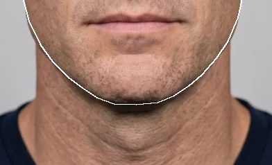
Before 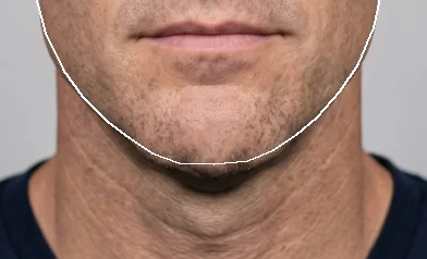
Right after+6.7 mm forward 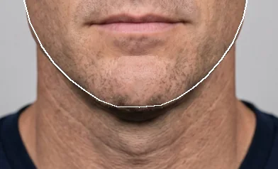
2 weeks+4.2 mm forward 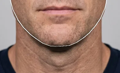
3 months+2.5 mm forward
Range at 3 months, from the spread in those studies
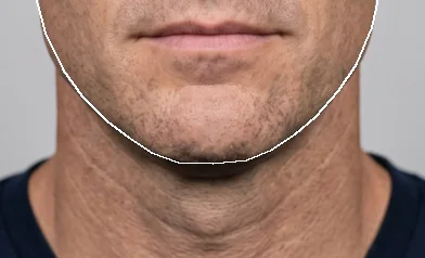
Low+1.2 mm Typical+2.5 mm 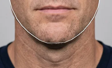
High+3.6 mm
In the app
Treatments are set in the units a clinic uses; the readouts and the 3D view come from the same numbers.
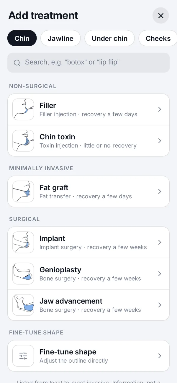
Treatments by area Toxin in units 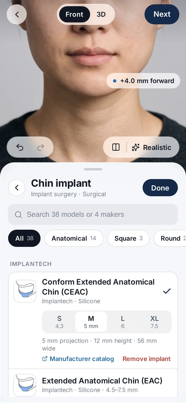
Implant models and sizes 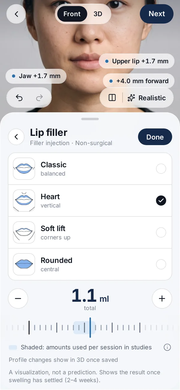
Lip filler in ml, by style 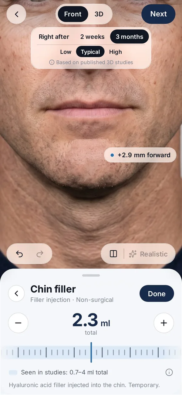
Filler over time (in testing) Approximate mm, before → after 3D view from the depth scan Original and visualization
Faces are synthetic. Images are visualizations, not predictions or treatment results.
06Limits
Where clinical systems do things Veylo doesn’t.
Capture in milliseconds
VECTRA captures in 2.0–3.5 ms[7, 8]. A phone scan takes about 40 seconds of guided head turns, so movement and expression matter more.
Years of validation
VECTRA has a body of independent validation studies[22]. Veylo has none of its own yet.
A clinician in the loop
In a clinic, the person shaping the simulation knows the anatomy and what a treatment can do. Veylo is used without a clinician and does not judge what is achievable.
Measuring real change
Clinical systems measure surface and volume change between visits[8]. Veylo visualizes a change; it does not measure a treatment outcome.
Breast and body
VECTRA, Crisalix and LifeViz also cover breast and body[7, 9, 20]. Veylo covers the face only.
Neighbouring areas and swelling
Real treatments can affect nearby areas and swell for weeks[39]. Veylo keeps other areas still by design and shows a settled result, so it shows neither.
A generative realistic view
The photographic render can vary slightly between runs and may alter small details. The 3D editor does not.
What we measure, and how we will validate it.
What Veylo measures today
- Scan
- A fused 3D surface from TrueDepth depth, with landmarks. Each capture is checked for light, distance, pose and expression, and views that don’t align are dropped.
- Edit
- The change in mm the engine applies for each treatment and amount, from ratios in published studies — for example chin implants, genioplasty and masseter toxin, which narrowed lower-face width at every dose tested[15, 16, 41].
- Readouts
- Approximate before → after distances on the visualization. They describe the picture, not a future result; smartphone depth is weakest on deep and irregular areas[29].
How we will validate it
- 1
- 2Visualization vs result
With partner clinics: consenting patients scanned before treatment and at fixed times after (for example 2 weeks and 3 months), with the treatment record — product, ml or mm, site. We compare the visualization with the measured result inside the treated area, and against two baselines: “no change” and “average change”.
- 3
- 4Publication
A bioethics committee opinion before data are collected for publication, explicit consent under GDPR Article 9[42], and the method published with the results — including where Veylo falls short.
A phone scan alone cannot verify a small filler: TrueDepth face volumes differed from VECTRA by 3.1 cc on average[2], more than a typical 1 ml lip treatment. Small injectables need a clinic 3D camera for validation.
Rules we follow, and why.
- No scores, no suggestions
Veylo never rates a face, ranks styles or says what suits you. Surgeons have written about the drawbacks of 3D simulation in consultations[43].
- Never shown as a result
UK advertising guidance expects before-and-after photos to be genuine and representative[44]. A Veylo image is never a treatment result.
- Not a medical device
Veylo doesn’t diagnose or plan treatment. EU guidance sets out when software becomes a medical device[45].
- Face data stays on the phone
Unless you share it, and it never goes to analytics[17].
References
Studies link to their DOI and PubMed record. Product pages were accessed on 7 October 2026. Charts on this page are our own drawings of the published numbers; no figures were copied.
- 1Rudy HL, Wake N, Yee J, Garfein ES, Tepper OM. Three-dimensional facial scanning at the fingertips of patients and surgeons: accuracy and precision testing of iPhone X three-dimensional scanner. Plastic and Reconstructive Surgery 146(6):1407–1417 (2020). doi.org/10.1097/PRS.0000000000007387 · PubMed 33234980
- 2Hartmann R, Weiherer M, Nieberle F, et al. Evaluating smartphone-based 3D imaging techniques for clinical application in oral and maxillofacial surgery: a comparative study with the Vectra M5. Oral and Maxillofacial Surgery 29(1):29 (2025). doi.org/10.1007/s10006-024-01322-2 · PubMed 39792225
- 3Atiyeh BS, Issa OB, Daoud FJ, Baajour JA. Preoperative 3-dimensional simulation in aesthetic surgery. Plastic and Reconstructive Surgery – Global Open 14(8):e7999 (2026). doi.org/10.1097/GOX.0000000000007999 · PubMed 42559587
- 4Kim JS. Changes in volume of lips in 3-dimensional analysis and projection of lips in sonography after injection of particle-type hyaluronic acid filler utilizing a 9-point injection technique. Aesthetic Surgery Journal Open Forum 6:ojae076 (2024). doi.org/10.1093/asjof/ojae076 · PubMed 39670216
- 5Davis HD, Mazzaferro D, Habarth-Morales TE, et al. A large prospective volumetric and patient-reported outcome analysis of hyaluronic acid facial fillers. Plastic and Reconstructive Surgery 156(4):550–559 (2025). doi.org/10.1097/PRS.0000000000012135 · PubMed 40178806
- 6Voytik M, Khan S, Broach RB, Percec I. Three-dimensional volumetric analysis of chin augmentation using hyaluronic acid filler: a prospective study. Plastic and Reconstructive Surgery online ahead of print (2026). doi.org/10.1097/PRS.0000000000013346 · PubMed 42485070
- 7Canfield Scientific. VECTRA XT 3D imaging system — product page and technical specifications. canfieldsci.com (accessed 7 Oct 2026). www.canfieldsci.com/imaging-systems/vectra-xt-3d-imaging-system
- 8Canfield Scientific. VECTRA H2 3D imaging system — product page and technical specifications. canfieldsci.com (accessed 7 Oct 2026). www.canfieldsci.com/imaging-systems/vectra-h2-3d-imaging-system
- 9Crisalix SA. Crisalix — 3D simulation for patients and surgeons (home page). crisalix.com (accessed 7 Oct 2026). www.crisalix.com/en
- 10Crisalix SA. Crisalix 3D simulator: plans and pricing. crisalix.com (accessed 7 Oct 2026). www.crisalix.com/en/prices
- 11Galderma. Galderma launches FACE by Galderma™, an aesthetic visualization tool that simulates injectable treatment results in real time (press release, 22 March 2023). galderma.com (accessed 7 Oct 2026). www.galderma.com/news/galderma-launches-face-galdermatm-cutting-edge-aesthetic-visualization-tool-simulates
- 12Ilyosbekov S. Localize, don’t beautify: client-side control of image-editing APIs for cosmetic surgery previews. arXiv preprint 2608.02841 (pilot study, not peer-reviewed) (2026). arxiv.org/abs/2608.02841
- 13Curless B, Levoy M. A volumetric method for building complex models from range images. Proceedings of SIGGRAPH ’96 303–312 (1996). doi.org/10.1145/237170.237269
- 14Newcombe RA, Izadi S, Hilliges O, et al. KinectFusion: real-time dense surface mapping and tracking. 10th IEEE International Symposium on Mixed and Augmented Reality (ISMAR) 127–136 (2011). doi.org/10.1109/ISMAR.2011.6092378
- 15Moenning JE, Wolford LM. Chin augmentation with various alloplastic materials: a comparative study. International Journal of Adult Orthodontics and Orthognathic Surgery 4(3):175–187 (1989). pubmed.ncbi.nlm.nih.gov/2561746
- 16San Miguel Moragas J, Oth O, Büttner M, et al. A systematic review on soft-to-hard tissue ratios in orthognathic surgery part II: chin procedures. Journal of Cranio-Maxillofacial Surgery 43(8):1530–1540 (2015). doi.org/10.1016/j.jcms.2015.07.032 · PubMed 26321067
- 17Websitters Sp. z o.o. Veylo privacy policy. getveylo.app (accessed 7 Oct 2026). getveylo.app/privacy/
- 18Google. Gemini API: image generation (documentation; SynthID watermark). ai.google.dev (accessed 7 Oct 2026). ai.google.dev/gemini-api/docs/image-generation
- 19QuantifiCare. LifeViz® Mini — 3D imaging system for the face. quantificare.com (accessed 7 Oct 2026). www.quantificare.com/3d-photography-systems-old/lifeviz-mini
- 20Remma (distributor). QuantifiCare LifeViz Infinity 3D photography system. remma.fr (accessed 7 Oct 2026). remma.fr/en/model/lifeviz-infinity-3d-photography-system
- 213dMD. 3dMD — 3D and 4D human capture (home page). 3dmd.com (accessed 7 Oct 2026). 3dmd.com
- 22De Stefani A, Barone M, Hatami Alamdari S, et al. Validation of Vectra 3D imaging systems: a review. International Journal of Environmental Research and Public Health 19(14):8820 (2022). doi.org/10.3390/ijerph19148820 · PubMed 35886670
- 23White JD, Ortega-Castrillon A, Virgo C, et al. Sources of variation in the 3dMDface and Vectra H1 3D facial imaging systems. Scientific Reports 10:4443 (2020). doi.org/10.1038/s41598-020-61333-3 · PubMed 32157192
- 24Almadori A, Speiser S, Ashby I, et al. Portable three-dimensional imaging to monitor small volume enhancement in face, vulva, and hand: a comparative study. Journal of Plastic, Reconstructive & Aesthetic Surgery 75:3574–3585 (2022). doi.org/10.1016/j.bjps.2022.04.042 · PubMed 35659734
- 25Liu J, Zhang C, Cai R, Yao Y, Zhao Z, Liao W. Accuracy of 3-dimensional stereophotogrammetry: comparison of the 3dMD and Bellus3D facial scanning systems with one another and with direct anthropometry. American Journal of Orthodontics and Dentofacial Orthopedics 160(6):862–871 (2021). doi.org/10.1016/j.ajodo.2021.04.020 · PubMed 34814981
- 26Knoedler S, Alfertshofer M, Simon S, et al. Turn your vision into reality — AI-powered pre-operative outcome simulation in rhinoplasty surgery. Aesthetic Plastic Surgery 48(23):4833–4838 (2024). doi.org/10.1007/s00266-024-04043-9 · PubMed 38777929
- 27Canfield Scientific. ViewMyConsult. canfieldsci.com (accessed 7 Oct 2026). www.canfieldsci.com/imaging-systems/view-my-consult
- 28App Store. Pody Plastic Surgery Simulator — App Store listing (description). apps.apple.com (accessed 7 Oct 2026). apps.apple.com/us/app/pody-plastic-surgery-simulator/id6740537390
- 29Luo Y, Zhao M, Lu J. Accuracy of smartphone-based three-dimensional facial scanning system: a systematic review. Aesthetic Plastic Surgery 48(21):4500–4512 (2024). doi.org/10.1007/s00266-024-04121-y · PubMed 38831068
- 30Kühlman DC, Almuzian M, Coppini C, Alzoubi EE. Accuracy (trueness and precision) of four tablet-based applications for three-dimensional facial scanning: an in-vitro study. Journal of Dentistry 135:104533 (2023). doi.org/10.1016/j.jdent.2023.104533 · PubMed 37149254
- 31Persing S, Timberlake A, Madari S, Steinbacher D. Three-dimensional imaging in rhinoplasty: a comparison of the simulated versus actual result. Aesthetic Plastic Surgery 42(5):1331–1335 (2018). doi.org/10.1007/s00266-018-1151-9 · PubMed 29789868
- 32Yamamichi K, Nakanishi Y, Chen CY. Three-dimensional simulation accuracy and patient satisfaction with rhinoplasty. Aesthetic Surgery Journal Open Forum 7:ojaf110 (2025). doi.org/10.1093/asjof/ojaf110 · PubMed 41048375
- 33Lorange E, Bouhadana G, Oiknine N, Luc M, Borsuk DE. 3-dimensional simulation for breast augmentation: does the software actually work?. Plastic and Reconstructive Surgery 158(1):56–65 (2025 (issue 2026)). doi.org/10.1097/PRS.0000000000012683 · PubMed 41344311
- 34Knoops PGM, Borghi A, Breakey RWF, et al. Three-dimensional soft tissue prediction in orthognathic surgery: a clinical comparison of Dolphin, ProPlan CMF, and probabilistic finite element modelling. International Journal of Oral and Maxillofacial Surgery 48(4):511–518 (2019). doi.org/10.1016/j.ijom.2018.10.008 · PubMed 30391090
- 35ter Horst R, van Weert H, Loonen T, et al. Three-dimensional virtual planning in mandibular advancement surgery: soft tissue prediction based on deep learning. Journal of Cranio-Maxillofacial Surgery 49(9):775–782 (2021). doi.org/10.1016/j.jcms.2021.04.001 · PubMed 33941437
- 36Berends B, Bielevelt F, Baan F, et al. Soft-tissue prediction based on 3D photographs for virtual surgery planning of orthognathic surgery. Computers in Biology and Medicine 194:110529 (2025). doi.org/10.1016/j.compbiomed.2025.110529 · PubMed 40505289
- 37El Sewify O, Gorgy A, Sylvain M, Legler J, Zammit D. Patient perspective on preoperative simulation in plastic surgery: a systematic review of patient-reported outcomes. Plastic Surgery (Oakville) online ahead of print (2026). doi.org/10.1177/22925503261478090 · PubMed 42682868
- 38Harris S, Michon A. Defining and measuring ‘lift’ in soft tissue filler-based facial rejuvenation: a critical review and proposed framework. JPRAS Open 51:646–656 (2026). doi.org/10.1016/j.jpra.2026.07.028 · PubMed 42620772
- 39van der Vlis M, Dentino KM, Vervloet B, Padwa BL. Postoperative swelling after orthognathic surgery: a prospective volumetric analysis. Journal of Oral and Maxillofacial Surgery 72(11):2241–2247 (2014). doi.org/10.1016/j.joms.2014.04.026 · PubMed 25236819
- 40Yassa A, Akhavan A, Ayad S, et al. Facial aesthetics in artificial intelligence: first investigation comparing results in a generative AI study. Eplasty 25:e13 (2025). pubmed.ncbi.nlm.nih.gov/40661090
- 41Liew S, Rivers JK, Humphrey S, et al. Improvement of lower facial shape after treatment with onabotulinumtoxinA: secondary results from a phase 2 dose escalation study. Plastic and Reconstructive Surgery 157(2):258–269 (2025). doi.org/10.1097/PRS.0000000000012345 · PubMed 40801407
- 42European Union. Regulation (EU) 2016/679 (GDPR), Article 9: processing of special categories of personal data. EUR-Lex (2016). eur-lex.europa.eu/eli/reg/2016/679/oj
- 43Montemurro P, Savani L, Toninello P. The dark side of 3D simulation in breast augmentation: how to use its advantages and avoid its drawbacks. Aesthetic Surgery Journal 45(4):NP129–NP131 (2025). doi.org/10.1093/asj/sjae245 · PubMed 39696997
- 44Advertising Standards Authority / CAP (UK). Before and after photos (advice online). asa.org.uk (accessed 7 Oct 2026). www.asa.org.uk/advice-online/before-and-after-photos.html
- 45Medical Device Coordination Group. MDCG 2019-11: Guidance on qualification and classification of software in Regulation (EU) 2017/745 (MDR) and 2017/746 (IVDR). European Commission (2019). health.ec.europa.eu/system/files/2020-09/md_mdcg_2019_11_guidance_en_0.pdf
Spotted an error or an outdated detail? Write to [email protected].